Pilot study ‘Topological Characterization of the Retinal Microvascular Network Visualized by a Portable Non-mydriatic Fundus Camera HORUS 200 in healthy young subjects” is under way at the Faculty of Medicine in Podgorica.
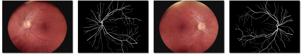
The microvascular system of the retina is an integral part of the microcirculation of the human body that can be directly studied in vivo in a simple and non-invasive way. The condition of these blood vessels can be recorded through digital photography. This approach gives us information not only about the health of the eye, but also the microcirculation throughout the entire body and it is used in the diagnosis of systemic diseases such as diabetes mellitus and hypertension.
Portable digital cameras are improving in quality and they are becoming more affordable. They are easy and quick to use, and do not require prior dilatation of the pupil, so they can be used in the primary health care setting. Also, this type of camera is light and can be used in the field for patients for whom health care is difficult to access. The digital photographs can then be easily transmitted for expert analysis by an ophthalmologist.
In order for this retinal imaging technology to be applied successfully in everyday practice, it is necessary that there is first a detailed description of the normal retinal anatomy visualized through this method.