Newest Trends in Development of Tools for Identification of Patients at Risk for Alzheimer Disease

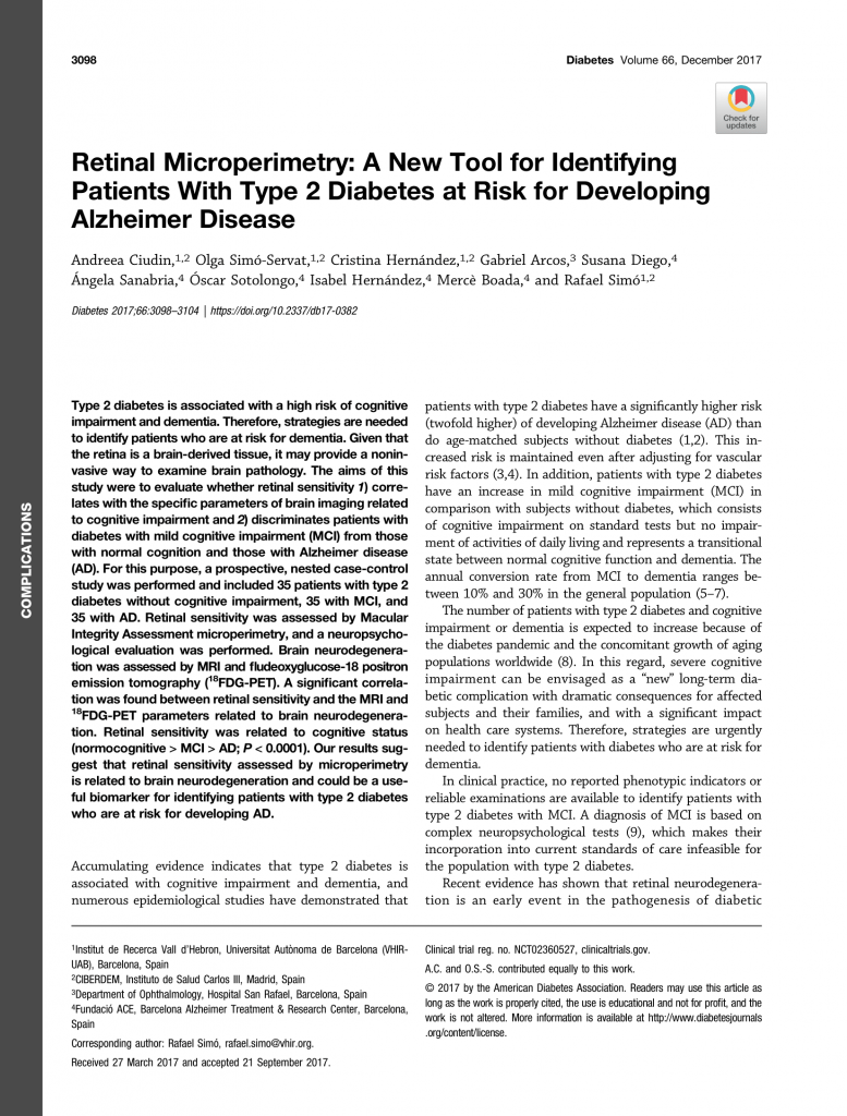
The paper “Retinal Microperimetry: A New Tool for Identifying Patients With Type 2 Diabetes at Risk for Developing Alzheimer Disease published by Ciudin et al in Diabetes in 2017 represents an important step forward in the development of non-invasive detection of patients at risk for the development of Alzheimer disease.

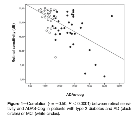
This is especially important in patients with type 2 diabetes, who have two-fold higher risk for developing dementia than age-matched people without diabetes, even after taking into account vascular risk factors. The paper demonstrated that retinal sensitivity assessed by microperimetry directly correlates with signs of brain neurodegeneration detected by MRI and FDG-PET. More importantly, retinal sensitivity significantly correlates with cognitive status of the patients.

These data suggest that retinal microperimetry could be applied as a new tool in identification of patients at risk for Alzheimer disease.

Read more at: https://doi.org/10.2337/db17-0382

Pilot Study – Use of Portable Non-mydriatic Fundus Camera

Pilot study ‘Topological Characterization of the Retinal Microvascular Network Visualized by a Portable Non-mydriatic Fundus Camera HORUS 200 in healthy young subjects” is under way at the Faculty of Medicine in Podgorica.

Image: Manual Segmentation of Retinal Microvascular Network

The microvascular system of the retina is an integral part of the microcirculation of the human body that can be directly studied in vivo in a simple and non-invasive way. The condition of these blood vessels can be recorded through digital photography. This approach gives us information not only about the health of the eye, but also the microcirculation throughout the entire body and it is used in the diagnosis of systemic diseases such as diabetes mellitus and hypertension.

Portable digital cameras are improving in quality and they are becoming more affordable. They are easy and quick to use, and do not require prior dilatation of the pupil, so they can be used in the primary health care setting. Also, this type of camera is light and can be used in the field for patients for whom health care is difficult to access. The digital photographs can then be easily transmitted for expert analysis by an ophthalmologist.

In order for this retinal imaging technology to be applied successfully in everyday practice, it is necessary that there is first a detailed description of the normal retinal anatomy visualized through this method.

The eye as a window to the brain – RECOGNISED project

“The eye as a window to the brain” – new European initiative, RECOGNISED, will determine the usefulness of the retina as a tool for identifying people with type 2 diabetes and cognitive impairment

An important EU-funded project has been launched to explore the biological pathways that may link the alterations observed in the retina with those present in the brain in people with type 2 diabetes (T2D).

Type 2 diabetes is known to be an independent risk factor for developing cognitive impairment and dementia, with studies showing that people living with T2D have a two-fold higher risk of developing Alzheimer’s disease (AD) when compared to the general population. AD is a neurodegenerative disease that leads to the progressive loss of brain cells, which causes cognitive decline and, eventually, dementia. People with cognitive impairment are more prone to have impaired diabetes self-management, poor glycaemic control and an increased incidence of diabetes-related complications, which presents significant challenges both for individuals and healthcare systems on how best to manage diabetes care. 

The four-year long RECOGNISED project will study the biological mechanisms that cause structural and functional alterations in the retina in people with type 2 diabetes, to determine whether these same pathways play a role in the events observed in the brain during the development of cognitive impairment and dementia. Importantly, RECOGNISED will reveal whether evaluating the retina, easily accessible with current non-invasive technologies, could help in identifying earlier cognitive impairment in people with T2D, so that appropriate support can be given. RECOGNISED will also analyse previously-collected data and samples from registries, cohorts and biobanks. By gaining knowledge on the mechanisms of disease, RECOGNISED will help to identify new potential therapeutic interventions.  

RECOGNISED brings together 21 partners from nine different countries, including academic institutions, small and medium enterprises (SMEs), the European infrastructure for translational medicine (EATRIS) and patient organisations, with complementary knowledge and expertise. RECOGNISED will receive almost €6 million in funding from the EU Horizon 2020 towards this programme with the final goal of improving the quality of life of people living with diabetes. In RECOGNISED, basic scientists and clinicians with extensive expertise in diabetes, ophthalmology and neurology will use state-or-the-art technologies to undertake the experimental and clinical studies that form part of this ambitious project.

Project Coordinator: Professor Rafael Simó, Vall d’Hebron Research Institute, Barcelona (Spain)

Project Partners: Queen’s University Belfast (UK), AIBILI (Portugal), Utrecht University Medical Center (Netherlands), Academic Medical Center, Amsterdam (Netherlands), Tor Vergata University of Rome (Italy), San Raffaele University Hospital (Italy), University of Milan (Italy), University of Southern Denmark (Denmark), University Hospital Trust, Charity of Novara (Italy), Fundacio Assistencial de Mutua de Terrassa (Spain), Catalan Health Institute (Spain), University of Montenegro (Montenegro), Clinical Center of Montenegro (Montenegro), University of Cadiz (Spain), EATRIS (Netherlands), Alzheimer Europe (Luxembourg), IDF-Europe (Belgium), Anaxomics Biotech (Spain), Oxurion (Belgium), Genesis Biomed (Spain), Vall d’Hebron Research Institute, Barcelona (Spain).

This project receives funding from the European Union’s Horizon 2020 research and innovation programme under grant agreement No 847749. This material reflects only the authors’ views, and the Commission is not liable for any use that may be made of the information contained therein.
Conference Paper Presentation: IT2020

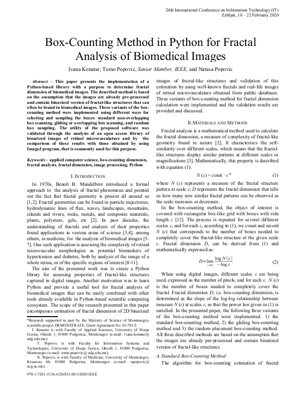
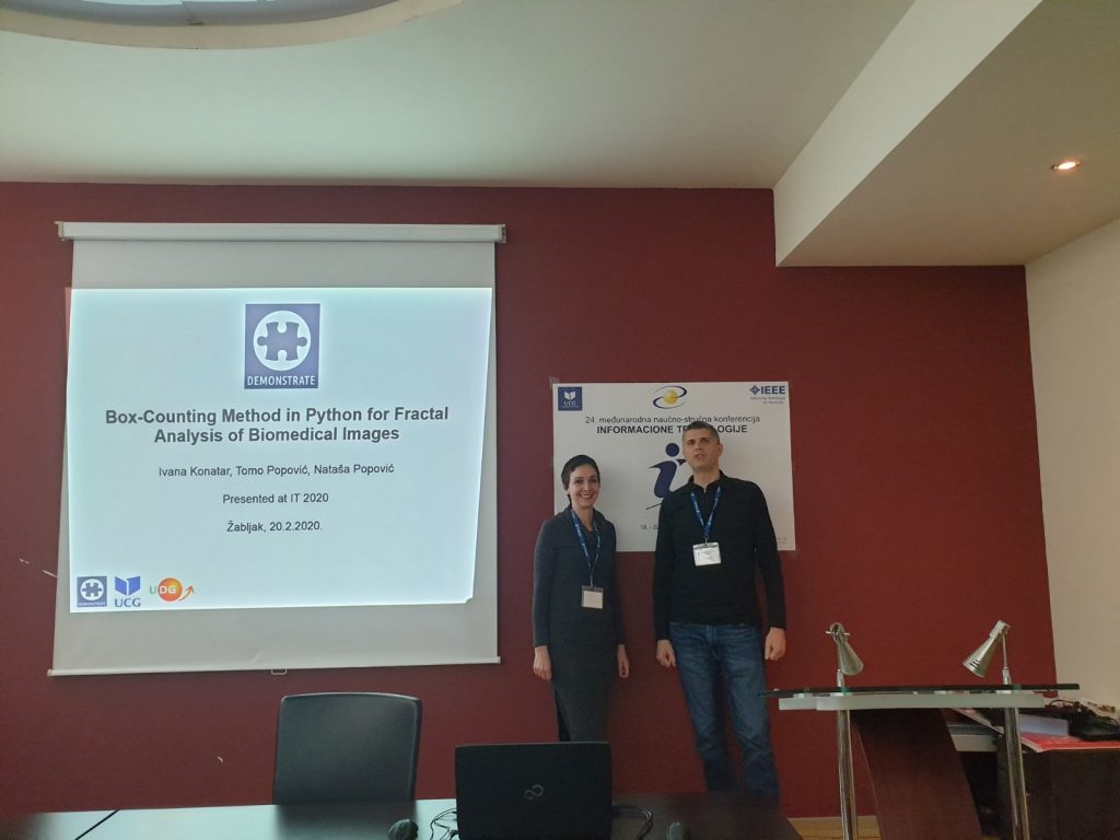
Project DEMONSTRATE was featured in the scientific paper “Box-counting method for fractal analysis of biomedical images” that was presented at the IEEE 24th International conference on Information Technology, IT2020 on 20. February, 2020. The paper discussed an implementation of a software library for fractal analysis aimed at quantification of complexity of microvascular network in human retina. The software calculates box-counting fractal dimension that can be used as a potential biomarker of various diseases. This research effort demonstrated that the use of this software module offers a very good flexibility in performing in silico experiments with digital images of retina and possibly other biomedical images. More info at: link.

Click to open in IEEE Xplore Library
At the IT2020 paper presentation session

Doctoral Thesis Application

The DEMONSTRATE project strives to engage young medical doctors who are enrolled in PhD studies at the Faculty of Medicine in Montenegro. The results of the project DEMONSTRATE will be an integral part of their doctoral dissertations.

On January 31st 2020, a young medical doctor, researcher, and PhD student participating in the DEMONSTRATE project, Isidora Rovčanin Dragović, submitted the Application for her Doctoral Thesis Topic. The title of the proposed research topic is: ”A new method for stratification of the risk for Alzheimer’s disease in patients in Montenegro”. The research plans to study the role of miRNA species as putative biomarkers of Alzheimer’s disease. With the help of the Italian scientists from IBIOM in Bari, she will work to acquire additional education and master practical skills necessary to accomplish the project goals.

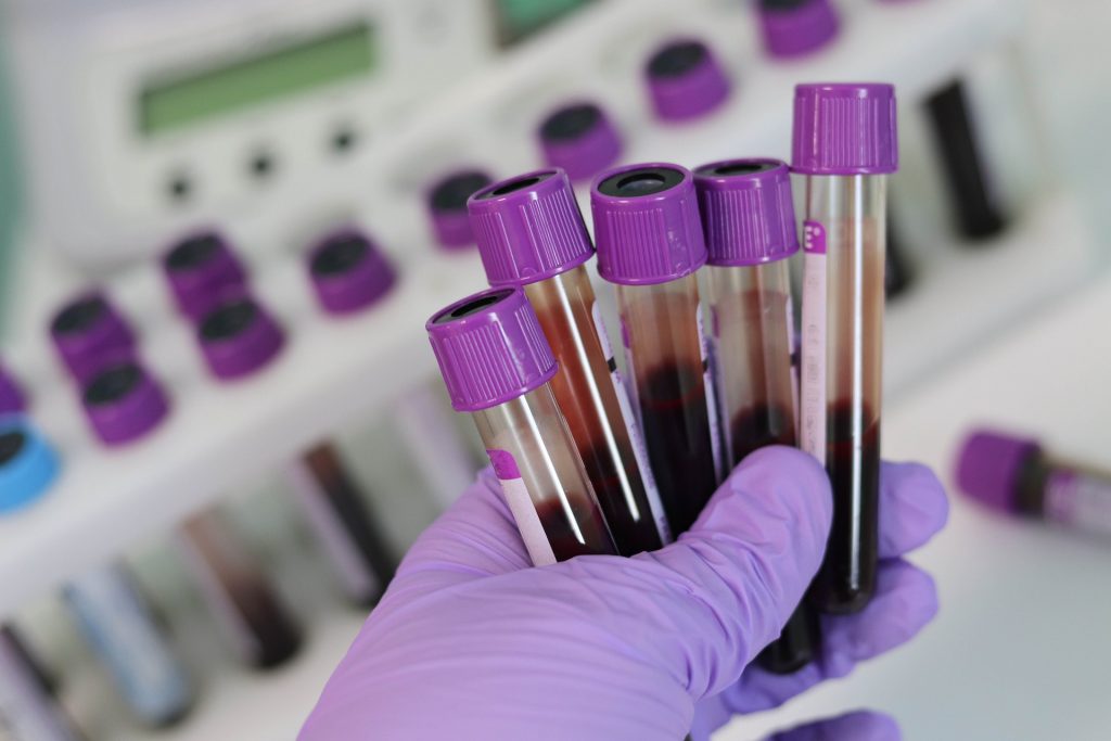
Image: Biological Samples (pixabay.com)

Visiting Lecture by Stela Vujosevic, MD, PhD

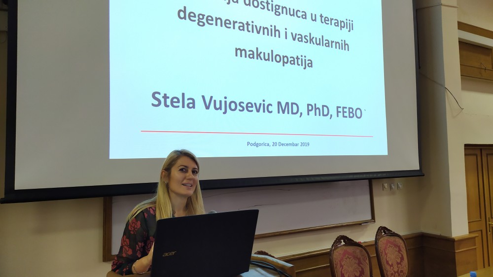
On December 20th, the Faculty of Medicine of the University of Montenegro was honored to host two invited lectures by Dr. Stela Vujošević, the expert adviser on the project DEMONSTRATE, from the Medical Center for Retina Diseases at the University Hospital “Maggiore della Caritá” in Novara, Italy.

Stela Vujo[ević (Image: UCG)

The lectures were titled:

  1. Recent developments in the treatment of degenerative and vascular maculopathies
  2. Application of non-invasive “imaging” biomarkers in the treatment of diabetic macular edema: the concept of precision medicine

This visit was organised by the Faculty of Medicine Vice Dean for Science and funded by the Ministry of Science of Montenegro, through the program of Guest Lecturers. The purpose of this program is to engage prominent scientists, university professors or entrepreneurs from the world to deliver intensive courses for students and / or scientific and academic staff at universities in our country, in the areas that are identified as a priority for the Smart Specialization Strategy.

Successful HORIZON 2020 Grant Application

The networking and collaboration established though the DEMONSTRATE project resulted in achieving a milestone in the area of sustainability and increasing the visibility of Montenegrin institutions in the field of medical research in the world.

The Faculty of Medicine of the University of Montenegro and the Clinical Center of Montenegro won the HORIZON 2020 grant as partner institutions on the project titled Unravelling the common pathways and identifying patients at risk for dementia – RECOGNISED. This is a multinational project involving 21 research institutions and teaching hospitals from Europe that are leaders in the field of research related to Alzheimer’s dementia and diabetes. The coordinating organization is Fundacio Hospital Universitari Vall D’Hebron/ Institut de Recerca (VHIR).

The goal of this project is to evaluate patients with diabetes type 2 who are at increased risk developing dementia based on microvascular or neurodegenerative changes we observe on the retina. This 4-year project will start on January 1st 2020. More information on the project is available at the following link.

(image: EC Horizion 2020)

Journal Paper Publication: Scientific Reports

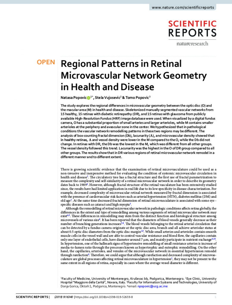
A scientific paper named “Regional Patterns in Retinal Microvascular Network Geometry in Health and Disease” (authored by N. Popovic, S. Vujošević, and T. Popovic) has been published in the Scientific Reports on November 8th 2019. This work is a result of a collaboration of scientists from the Faculty of Medicine of the University of Montenegro, the Faculty for Information Systems and Technologijes, University of Donja Gorica from Podgorica and the Eye Clinic, University Hospital “Maggiore della Caritá” from Novara, Italy. This research was conducted to develop a methodological approach that will also be used to analyze the fundus photographs that we will collect through the DEMONSTRATE project. More information at the following link.

Click on image to read the paper

Semi-Annual Progress Meeting

On October 2nd, the Faculty of Medicine in Podgorica hosted the semi-annual meeting for the project “New methods for risk stratification for the progression of cancer and Alzheimer’s disease in patients in Montenegro – DEMONSTRATE”. In addition to the team members from the Clinical Center of Montenegro from Non-governmental Organization Futura, the members of the Institute of Biomembranes, Bioenergetics and Molecular Biotechnology from Bari and an external expert from University Hospital Maggiore della Carita’ from Novara, Italy, participated in the meeting.

Semi-annual Progress Meeting for DEMONSTRATE project in Podgorica